March 13, 2019 (HealthDay News)
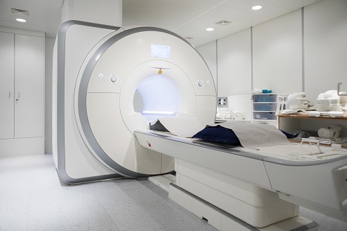
For patients with multiple sclerosis (MS), use of a contrast-based agent at follow-up magnetic resonance imaging (MRI) does not change the diagnosis of interval disease progression, according to a study published online March 12 in Radiology.
Paul Eichinger, M.D., from the Technische Universität München in Germany, and colleagues conducted a retrospective study based on a local prospective observation cohort to examine whether use of contrast material impacts the detection of new or enlarged MS lesions. A total of 507 follow-up MRIs from 359 patients with MS were assessed.
The researchers found that 264 of the images showed interval progression, with a total of 1,992 new or enlarged and 207 contrast-enhancing lesions. Four of these lesions were detected retrospectively on only the nonenhanced images, representing 1.9 percent of the enhancing lesions and 0.2 percent of new or enlarged images. On fluid-attenuated inversion recovery (FLAIR)-based subtraction maps, nine enhancing lesions were not detected (0.6 percent of 1,442). Contrast-enhanced sequences did not reveal interval progression that was missed in the readouts of the nonenhanced sequences with use of either double inversion recovery- or FLAIR-based subtraction maps in any of the 507 images.
“Our data do not support the notion that the use of contrast material results in higher sensitivity for the detection of new or enlarged lesions in MS compared with unenhanced MRI at 3.0 T,” the authors write.
Several authors disclosed financial ties to the biopharmaceutical and health care industries.
Copyright © 2019 HealthDay. All rights reserved.